Carotid Doppler: Ultrasound of the Neck Vessels

The carotid arteries play a central role in blood supply to the brain. The Cardiological diagnostics uses the carotid Doppler to measure blood flow in vessels using ultrasound and to detect narrowing and circulatory disorders at an early stage. The next step is Duplex ultrasonography, which combines Doppler ultrasound with a two-dimensional B-mode ultrasound image. This also makes it possible to visualise the structure of the vessel wall, plaques or constrictions.

Dr Bernhard Nagel examines a patient using ultrasound.

Indications for carotid Doppler

Doppler ultrasonography is suitable for examining a wide variety of blood vessels.

The carotid Doppler is used, among other things, when there is suspicion of:

  • Arteriosclerosis
  • Hypertension

Further possible indications include stroke prophylaxis, visual disturbances not originating from the eye itself, and syncope without cardiological findings.

Risk factors for vascular changes

A vessel Doppler is particularly useful for risk groups with specific pre-existing conditions that are considered risk factors for vascular changes.

As a risk group for vascular changes, consider individuals with:

  • Hypertension (high blood pressure)
  • Diabetes Mellitus
  • Raised cholesterol (high blood fat levels)
  • Previous stroke
  • Transient ischaemic attack

There are no contraindications for carotid Doppler. However, diagnostic precision can be impaired by limited acoustic windows (e.g., in cases of massive calcifications or anatomical peculiarities).

Findings and Diagnostic Goals of Carotid Doppler

The focus is on the detection of vascular wall thickening, cholesterol plaques (deposits) and dissections (tears in the vessel wall). Furthermore, Doppler measurement allows for the visualisation and acoustic representation of blood flow, including the assessment of flow direction, speed and potential accelerations and turbulence.

Additionally, a carotid Doppler can be used to assess whether both carotid arteries are equally perfused or if there are differences between the right and left sides. The surface of the vessel wall also becomes visible: it can appear smooth or irregular, which may indicate structural changes. If findings are already known, a carotid Doppler also allows for an assessment of whether the condition of the vessels has changed over time. This provides a differentiated picture of the structure and function of the carotid arteries and their blood flow.

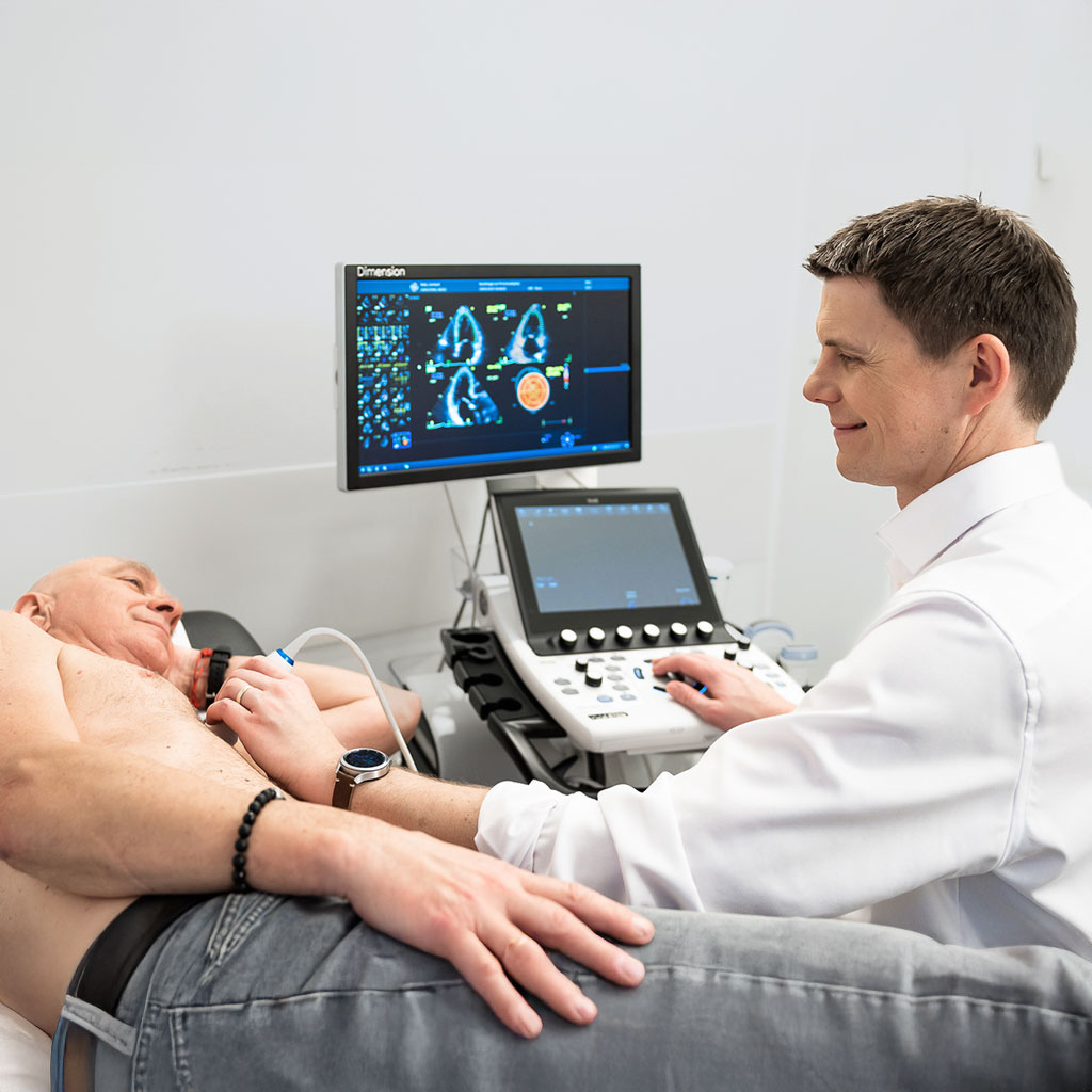
The carotid Doppler is a proven, gentle ultrasound examination of the neck vessels and is part of routine vascular diagnostics. A small, non-invasive ultrasound device, which displays live images on a monitor, is used for the examination.

Carotid Doppler: Functions and Technology

A carotid Doppler is one of the non-invasive routine examinations in vascular diagnostics. The Doppler is considered a reliable and gentle method for imaging the neck vessels.

Depending on the question, different Doppler methods are used:

  • Colour duplex Doppler: shows the vessel wall and visualises blood flow in colour.
  • Pulsed-Wave Doppler: precisely measures flow velocity at a defined location.
  • Continuous-Wave Doppler: is particularly suitable for the precise measurement of very high flow velocities in cases of severe vascular stenosis.

This allows for a detailed assessment of vascular structure and blood flow.

Carotid Doppler ultrasound procedure

A carotid Doppler is performed in the treatment room and carried out by the doctor. The patient sits slightly elevated while the physician scans the left and right sides of the neck with a small ultrasound probe. A gel ensures optimal skin contact and clear images. It is important to sit still and not to speak.

The results will then be discussed and a written report will be drawn up. If necessary, treatment will be carried out in the course of time. Depending on the findings, a follow-up examination of the vessels, usually once a year, may be advisable. Doppler ultrasonography is uncomplicated. It requires no preparation, only takes a few minutes, and allows an immediate return to everyday activities. It is only recommended that you do not wear a necklace during the carotid Doppler.

Carotid Doppler: Cost covered by health insurance

Statutory health insurance providers cover carotid artery examinations if they are medically necessary, for example, in cases of symptoms, abnormal findings, or pre-existing conditions. In these instances, a referral from a GP is required. However, if a carotid Doppler examination is carried out purely for preventative reasons without a specific indication, it usually needs to be paid for privately as an individual health service (IGeL).

Private health insurance companies generally reimburse the carotid examination, as it can be billed according to the German Medical Fee Schedule (GOÄ). The exact conditions may vary depending on the policy, so it is worth consulting in advance. Have you received a referral for a carotid Doppler and would like to make an appointment at our cardiology practice? Give us a call or use our online appointment booking system.

Important questions about carotid Doppler

The Doppler scan is non-invasive and painless. The gel may feel cold.

The Doppler ultrasound takes only about five minutes.

Immediately after the examination. The images can be assessed straight away and the result can be discussed directly with patients.

There are no contraindications for the Doppler Munich.

We use the Munich Doppler to check blood flow in the neck vessels (carotids) for instance with suspected arteriosclerosis, arterial hypertension, or after a stroke.